
Eye Anatomy Terms Defined
Have you ever been curious about the inner workings of your eyes? Let’s look into the human eye and learn about some of the key players responsible for its function.

Sclera
the armor.
The outer coating of the eye, giving the eyeball its opaque white hue.
Conjunctiva
the window trim & shutters.
A thin translucent membrane layer that covers the eye and insides of the eyelids. It also produces mucous and tears to keep the eyes lubricated.
This is the site of bacterial, viral, and allergic infections of the eye.
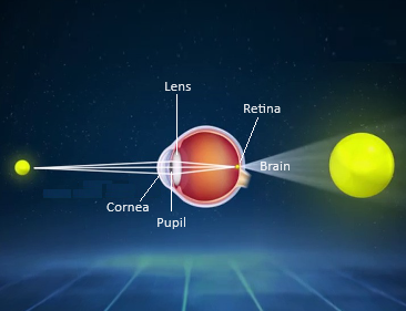
Cornea
the gatekeeper & the glass window.
A clear protective surface tissue that encases the front of the eye. Together with the sclera, it helps protect the eye by keeping out dirt, germs and other debris, while also filtering out some UV rays.
The eye’s focusing system starts here as light enters through the cornea.
Iris
the blinds & shades.
The colored part of the eyes that controls the size of the pupil and the amount of light reaching the retina.
The darker the environment, the larger the iris muscles dilate the pupils to allow more light and enhance vision. The brighter the environment, the more the iris muscles constrict the pupils to allow less light inside and reduce glare.
Pupil
The aperture.
The opening at the center of the iris through which light passes.

Lens
the camera.
The crystalline core of the eye that focuses light from the pupil to the retina and switches depth of focus from far to near distances.
This is the part of the eye that is susceptible to cataracts - The lens begins to cloud overtime. While performing cataract surgery, the clouded lens is replaced with an artificial intraocular lens (IOL).
Retina
The interior fortified wall.
The layer of cells lining the back wall inside the eye. The retina captures the focused light from the lens and sends signals to the brain to perceive an image — OUR VISION.
This section of the eye is susceptible to holes, tears, or a detachment, causing visual disturbances or abnormal floaters/light flashes in our vision.
Retinal Vessels
The fuel.
Vasculature that provides proper blood flow and circulates oxygen to and from the retinal layers. The arteries supply nutrients to the retina, while the veins exit the retina by coursing through the optic nerve toward the heart.
The retinal arteries and veins are susceptible to blockages leading to partial vision loss from uncontrolled systemic conditions such as cholesterol, blood pressure, and diabetes.
Vitreous Humor
The shock absorber.
A gel-like substance that fills the back of the eye. It keeps all contents of the retina secure when the body undergoes a sudden movement or physical injury.
This is the site where the common “safe” floaters form when parts of its gel fibers begin to separate from the whole vitreous overtime.
Optic Nerve
the network.
The 2nd cranial nerve that connects the eyes to the brain. The optic nerve is a bundle of >1 million nerve fibers that send chemical and electrical messages to the brain to conceive vision.
The location of the optic nerve is also where our physiological blind spot is.
This is the part of the eye damaged from neuropathies such as glaucoma and neuritis from multiple sclerosis (MS).
Macula
the capital.
The central region of the retina that provides the sharpest, fine-detailed vision and interprets color at its focal point - the Fovea. It is our central vision.
This is the part of the retina susceptible to vision degradation from age-related macular degeneration and complications of diabetes leading to macular edema or neovascularization.
